


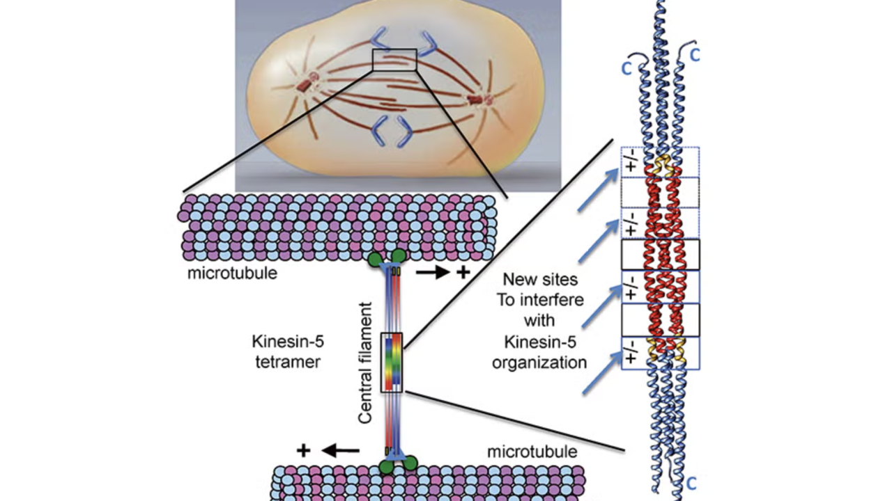
Structural Basis for the Assembly of the Mitotic Motor Kinesin-5 into Bipolar Tetramers
During cell division, an organized and symmetric intracellular apparatus, named the mitotic spindle, composed of microtubules (and associated molecular machines), coordinate the accurate alignment and equal distribution of newly duplicated chromosomes into two new cells. When the organization of mitotic spindles becomes mis-regulated, a variety of human disease conditions can result, including early onset cancers and human birth defects. Our laboratory studies the molecular mechanisms of microtubule dynamics and organization during mitotic cell division.
Molecular motors in living cells bind and generate motile forces through “step-by-step walking” along microtubules. These motor activities move chromosomes and mechanically orchestrate events during mitotic cell division. We are focused on an evolutionarily well-conserved molecular motor class, Kinesin-5, which are fundamentally responsible for elongating mitotic spindles to form two new cells. The organization of mitotic motor kinesin-5 is unique from all other molecular motors in that they form elongated bipolar molecular assemblies, composed of four subunits, with two sets of active motile motors pointed towards opposite ends of a rigid central rod. The central rod is critical for transmitting forces between the motile ends. The “dual-headed” arrangement of kinesin-5 is critical for its function as it preferentially binds microtubules emanating from opposite poles of the mitotic spindle and mediates their anti-parallel sliding near the end of the cell division cycle.
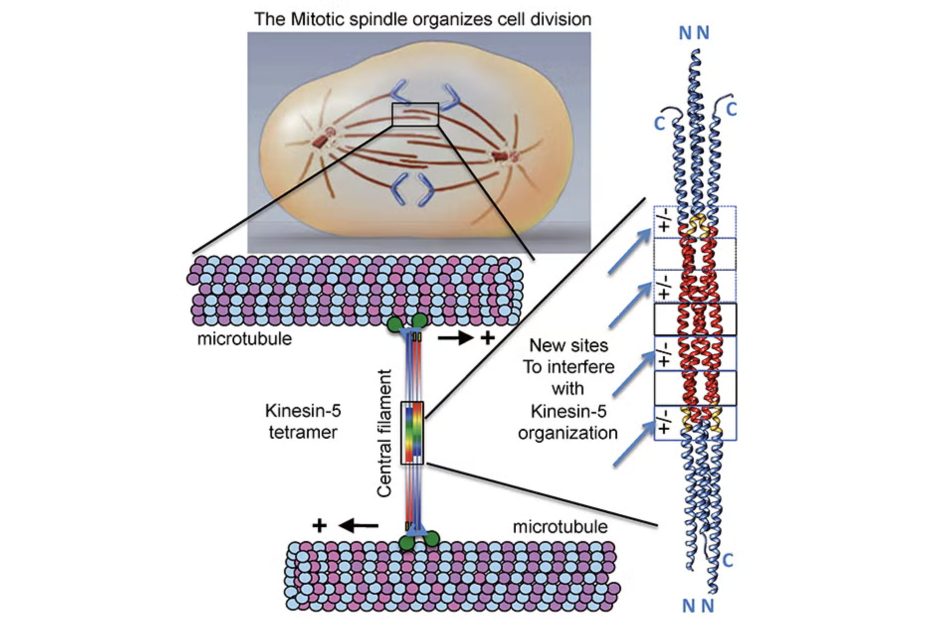
Our laboratory made a major breakthrough, in collaboration with Jonathan Scholey’s group at UC-Davis, in determining the atomic three-dimensional structure four-subunit kinesin-5 central rod region, which specifies the mitotic kinesin-5 organization into “dual headed” bipolar molecular machines. We discovered a precise three-dimensional organization where four kinesin-5 subunits form bipolar assemblies. We observed an alternating pattern of seven symmetrically organized interface elements composed of uniquely interacting amino acids. Four out of seven of these interface elements are ionic in nature and form exposed “molecular pockets” that are accessible to small chemical molecules from the solution. Mutations of the ionic interacting amino acids severely disrupt the kinesin-5 four-subunit organization, leading misassembled kinesin-5 molecules. The kinesin-5 structure suggests a novel strategy in targeting kinesin-5 inactivation that is conceptually more effective in controlling mitotic spindle elongation than the previous strategies as it is specifically focused on disrupting kinesin-5 assembly by disassembling its central bipolar rod filament, the main region that transmits force between the two kinesin-5 motile ends.
Published in eLife.