News

Structural basis of kinesin-1 autoinhibition and its control of microtubule-based motility

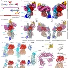
Kinesin-1, a heterotetramer of two kinesin heavy chains (KHC) and two kinesin light chains (KLC), drives microtubule-based transport of organelles and macromolecules, and its dysfunction is linked to ALS and hereditary spastic paraplegia. In the absence of cargo or activating factors, it adopts a compact autoinhibited conformation, but the molecule’s length and flexibility had prevented structural characterization of this state. We determined the first cryo-EM structure of the full-length autoinhibited kinesin-1 heterotetramer.

A unified mechanism for tubulin cofactors catalyzing α/β-tubulin biogenesis and degradation

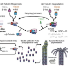
We solved a decades-old mystery in cell biology by revealing how a group of proteins called tubulin cofactors build and dismantle the molecular building blocks of the cell’s internal skeleton. Using cryo-electron microscopy, we captured this molecular machine frozen at each stage of its cycle, showing precisely how it malfunctions in children with rare neurological diseases such as infantile encephalopathy and corpus callosum hypoplasia.

Cryo-EM structures of the tubulin cofactors reveal the molecular basis of alpha/beta-tubulin biogenesis

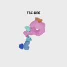
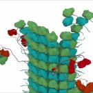
We determined the first cryo-EM structures of the tubulin cofactor complex TBC-DEG bound to an αβ-tubulin heterodimer, revealing for the first time how TBCD, TBCE, and the Arl2 GTPase form a cage-like assembly that encircles and stabilizes the αβ-tubulin dimer to regulate its biogenesis. TBC-DEG wraps around β-tubulin while TBCE extends along α-tubulin, and a companion structure with TBCC shows that TBCC engages Arl2 and TBCD to contact the αβ-tubulin intradimer interface, promoting TBCE rotation while TBCD holds β-tubulin.

Cryo-EM structures of plant Augmin reveal coiled-coil assembly, antiparallel dimerization, and NEDD1 binding

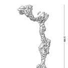
Microtubule branch nucleation, which builds complex microtubule networks in plants and metazoans, depends on the Augmin complex and NEDD1 to recruit and activate the γ-tubulin ring complex (γ-TuRC). Combining reconstitution, crosslinking mass spectrometry, and 3.7–7.3 Å cryo-EM, we built a complete de novo model of Arabidopsis Augmin, revealing long-range multi-coiled-coil interfaces that stabilize its 40-nm hetero-octameric fork-shaped architecture.


The kinesin-5 tail domain directly modulates the mechanochemical cycle of the motor domain for anti-parallel microtubule sliding

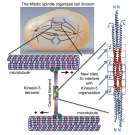
Kinesin-5 motors organize mitotic spindles by sliding apart microtubules. They are homotetramers with dimeric motor and tail domains at both ends of a bipolar minifilament. Here, we describe a regulatory mechanism involving direct binding between tail and motor domains and its fundamental role in microtubule sliding. Kinesin-5 tails decrease microtubule-stimulated ATP-hydrolysis by specifically engaging motor domains in the nucleotide-free or ADP states. Cryo-EM reveals that tail binding stabilizes an open motor domain ATP-active site.

Microtubule polymerase and processive plus-end tracking functions originate from distinct features within TOG domain arrays

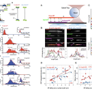
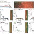
Microtubule polymerases accelerates tubulin polymerization while processively tracking microtubule (MT) plus ends via tumor overexpressed gene (TOG) domain arrays. We describe a relationship between MT plus-end tracking and polymerase functions revealing their distinct origin within TOG arrays using structural based model-based mutants, microtubule dynamic assays, single molecule fluorescence and studies of the mutants within fission yeast cells. We study Alp14 mutants designed based on structural models, with defects in either tubulin recruitment or self-organization.

Structural basis of tubulin recruitment and assembly by microtubule polymerases with tumor overexpressed gene (TOG) domain arrays


TOG (tumor overexpressed gene) domain arrays, found in microtubule polymerases such as yeast Alp14, organize and then polymerize αβ-tubulin at microtubule plus ends. Binding assays showed that TOG1 binds tubulin more strongly than TOG2, and a crystal structure of a Saccharomyces kluyveri Alp14 homodimer revealed two TOG1 and two TOG2 domains, each holding a tubulin dimer, arranged in a square with TOGs inside and tubulin outside.

Tubulin cofactors and Arl2 form a cage-like chaperone that catalyzes αβ-tubulin assembly through GTP hydrolysis: Reconstitution of new molecular machine

The building block of microtubules is composed of two proteins called α-tubulin and ß-tubulin which are assembled into a single polarized αß-tubulin subunit. Microtubules are very dynamic structures that can rapidly change length as individual αβ-tubulin units are either added or removed to their growing or shrinking ends. The assembly of aß-tubulin has for long remained a mystery despite extensive genetic and cell biology studies. Several proteins known as tubulin cofactors and an enzyme called Arl2 help to build a vast pool of αβ-tubulin that are able attach to the microtubules.

Structural Basis for the Assembly of the Mitotic Motor Kinesin-5 into Bipolar Tetramers

During cell division, an organized and symmetric intracellular apparatus, named the mitotic spindle, composed of microtubules (and associated molecular machines), coordinate the accurate alignment and equal distribution of newly duplicated chromosomes into two new cells. When the organization of mitotic spindles becomes mis-regulated, a variety of human disease conditions can result, including early onset cancers and human birth defects. Our laboratory studies the molecular mechanisms of microtubule dynamics and organization during mitotic cell division.